The two-tier motor circuit, and how it breaks
Last reviewed November 16, 2026. OwieClear editorial team. Waiting for clinician review. Not medical advice. Talk with your doctor about your own care.
Moving a finger takes two nerve cells arranged end to end: one from the brain to the spinal cord, one from the spinal cord to the muscle. Between them they can span a metre.
What makes ALS distinctive among neurological diseases is that it attacks both tiers at once, and the combination of signs that produces is what allows a diagnosis to be made at all.
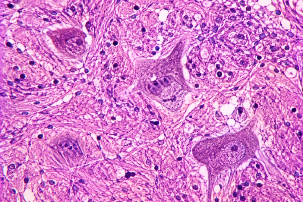
Related pages: ALS (amyotrophic lateral sclerosis)
The bottom line
- Upper motor neurons run from the motor cortex to the spinal cord.
- Lower motor neurons run from the spinal cord or brainstem to muscle.
- Upper motor neuron damage causes stiffness and overactive reflexes; lower motor neuron damage causes wasting and twitching.
- Degeneration usually begins at the far end, at the junction with muscle, rather than at the cell body.
- Why both tiers are affected in ALS, when most diseases hit one or the other.
- What determines where in the body the process starts.
The story, step by step
What the evidence labels mean
Brain to spinal cord
What happens
The upper motor neuron starts in the motor cortex and runs down through the brain and into the spinal cord.
What the research shows
- Its long axon travels in the corticospinal tract, through the lateral part of the spinal cord. That tract is the lateral in amyotrophic lateral sclerosis.
- Its job is not only to command movement but to restrain the spinal reflexes below it.
- When upper motor neurons degenerate, that restraint is lost: muscles become stiff and tight, a condition called spasticity.
- Reflexes become brisk and overactive, and abnormal reflexes appear, including the Babinski sign, where the big toe moves upward when the sole is stroked.
- Speech can become slow, strained and effortful, which is a distinct pattern from the slurring of lower motor neuron damage.
Why it matters
Brisk reflexes in a limb that is also wasting is an unusual combination, and it is one of the strongest pointers to ALS.
What's still uncertain
- Spasticity varies a great deal between people and is sometimes minimal.
Read more
Next: Tier two
Spinal cord to muscle
What happens
The lower motor neuron sits in the anterior horn of the spinal cord, or in the brainstem for head and neck muscles.
What the research shows
- Its axon runs out to the muscle and ends at the neuromuscular junction, where the signal is passed to the muscle fibre.
- One motor neuron supplies many muscle fibres, and together they are called a motor unit.
- When a lower motor neuron dies, its entire motor unit loses its signal. The muscle fibres waste, which is the visible atrophy.
- Dying motor neurons become electrically unstable, firing on their own. That produces fasciculations, the visible rippling twitch under the skin.
- Cramps are common for the same reason, and often appear early.
Why it matters
Twitching and wasting together, in the same muscle, is the lower motor neuron signature. Twitching alone, in a strong muscle, usually is not.
What's still uncertain
- Fasciculations occur in many healthy people and in several other conditions.
Read more
- NINDS: Amyotrophic lateral sclerosis (ALS)Government
- OwieClear: How the connection to muscle is lostOwieClear
Next: Both at once
The pattern that makes the diagnosis
What happens
Most neurological conditions damage one tier. ALS damages both, and that is diagnostically decisive.
What the research shows
- A wasted, twitching muscle with brisk reflexes is the combination, and it is unusual.
- A neurologist looks for both sets of signs in several regions of the body: bulbar, cervical, thoracic and lumbosacral.
- Diagnostic criteria, successively the El Escorial, Awaji and now Gold Coast criteria, formalize how many regions must show these signs.
- Conditions affecting only one tier exist, including primary lateral sclerosis, upper only, and progressive muscular atrophy, lower only. Both progress more slowly than typical ALS.
- Electrical testing can detect lower motor neuron loss in muscles that still look and feel normal, which is how the diagnosis is extended beyond what is visible.
Why it matters
This is why diagnosis requires a neurologist and electrical testing rather than a scan. The pattern is what matters, not any single finding.
What's still uncertain
- Early in the disease the full pattern may not yet be present, which is a common reason for diagnostic delay.
Read more
- NINDS: Amyotrophic lateral sclerosis (ALS)Government
- OwieClear: How ALS is diagnosedOwieClear
Next: Dying back
Failing at the far end first
What happens
The degeneration does not begin where most people assume.
What the research shows
- Motor neurons are among the largest cells in the body. A single cell can run from the lower spinal cord to a muscle in the foot.
- That cell must transport mitochondria, proteins and signalling molecules along the whole length of the axon.
- Evidence indicates that the connection at the neuromuscular junction is lost first, with the axon retracting back toward the cell body over time.
- This is called dying-back degeneration, and it means the earliest damage happens furthest from the cell's control centre.
- It is one reason treatments that protect the cell body may still fail to preserve function: the connection at the far end has already gone.
Why it matters
The disease starts at the handshake between nerve and muscle, not at the nerve cell itself, which is a significant finding for how treatment is approached.
What's still uncertain
- Most dying-back evidence comes from animal models, and how precisely it maps onto human ALS is debated.
Read more
- NINDS: Amyotrophic lateral sclerosis (ALS)Government
- OwieClear: The four mechanisms killing motor neuronsOwieClear
Next: Compensation
Why weakness appears late
What happens
The body hides this disease for a long time, which is why diagnosis is so often delayed.
What the research shows
- When a motor neuron dies, neighbouring surviving neurons sprout new branches and adopt its orphaned muscle fibres.
- This reinnervation maintains strength remarkably well, and a muscle can lose a substantial proportion of its motor neurons before weakness becomes noticeable.
- The surviving motor units become much larger than normal, which electrical testing can detect directly.
- Once compensation is exhausted, weakness can appear relatively suddenly, even though the underlying loss has been going on for a long time.
- This is why symptoms often seem to start abruptly in a disease that has in fact been progressing silently.
Why it matters
Weakness appearing suddenly does not mean the disease started suddenly. It means the compensation ran out.
What's still uncertain
- Estimates of how much motor neuron loss precedes symptoms vary and are difficult to measure in living people.